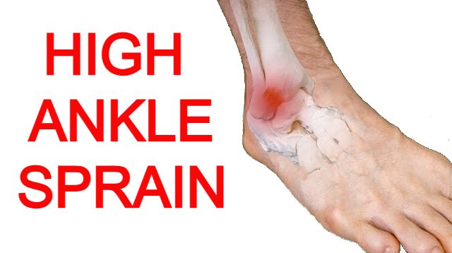
High Ankle Sprain
Understanding the High Ankle Sprain
Your ankle joint is incredibly well-designed. It allows for a wide range of movement and handles the stress of walking and other daily activities. You've probably heard of a typical ankle sprain. It's when you roll your ankle and damage the strong tissues (called ligaments) on the outside.
But there's another type of ankle injury, the high ankle sprain, which happens less often but can be more serious.
Unlike its lateral counterpart, a high ankle sprain affects the structures above the ankle joint itself, specifically the syndesmosis – the fibrous connection between the tibia and fibula bones in the lower leg. This seemingly subtle difference in location results in a distinct injury pattern, often requiring a longer recovery period and potentially leading to more significant long-term consequences if not properly managed.
Anatomy of a High Ankle Sprain
To truly understand the intricacies of a high ankle sprain, we must delve into the relevant anatomy. The syndesmosis is not merely a passive connection; it plays a crucial role in maintaining the stability of the ankle mortise, the bony arch formed by the tibia and fibula that cradles the talus bone. This tight articulation is essential for the efficient transfer of forces during activities like walking, running, and jumping.
The high ankle ligaments that constitute the syndesmosis are the anterior inferior tibiofibular ligament (AITFL), the posterior inferior tibiofibular ligament (PITFL), the interosseous membrane (IOM), and the transverse tibiofibular ligament (TTFL).
The AITFL, located on the front of the ankle, and the PITFL, situated at the back, are the strongest and most commonly injured components. The IOM is a strong, fibrous sheet that extends upwards between the tibia and fibula, providing further stability. The TTFL, a smaller ligament, runs transversely behind the PITFL.
Together, these structures ensure that the tibia and fibula remain tightly bound, limits external rotation, and allows for the smooth articulation of the ankle joint.
How Does a High Ankle Sprain Occur
The mechanism of injury in a high ankle sprain differs significantly from that of a lateral ankle sprain. While lateral sprains typically occur due to an inversion force (rolling the ankle inwards), a high ankle sprain occurs with a forceful external rotation of the foot relative to the lower leg, often combined with dorsiflexion (bending the foot upwards). Imagine planting your foot firmly and then having your body twist outwards, or experiencing a sudden, forceful upward bending of the ankle.
These scenarios can place tremendous stress on the syndesmotic ligaments, leading to stretching, partial tearing, or complete rupture. High-impact sports involving rapid changes in direction, such as soccer, football, basketball, and skiing, carry a higher risk of this type of injury. Direct trauma to the lower leg, such as a forceful blow, can also occasionally result in a high ankle sprain (syndesmotic injury).
High Ankle Sprain Diagnosis
The diagnosis of a high ankle sprain includes a thorough clinical evaluation and often involves the use of imaging studies. During the physical examination, a healthcare professional will carefully palpate the area above the ankle joint, specifically along the course of the syndesmotic ligaments. Tenderness in this region is a key indicator of a high ankle sprain.
Several specific provocative tests are also commonly performed to assess the integrity of the syndesmosis:
The squeeze test involves compressing the tibia and fibula together above the midpoint of the calf. Pain in the distal syndesmosis during this maneuver suggests injury and possible deltoid ligament involvement.
The external rotation test is performed by stabilizing the lower leg and passively externally rotating the foot. Pain in the syndesmotic area with this movement is highly suggestive of a high ankle sprain.
The dorsiflexion-abduction test involves maximally dorsiflexing and abducting the foot, which can also elicit pain if the syndesmosis is injured.
The fibular translation test assesses the anterior-posterior stability of the fibula relative to the tibia. Excessive movement or pain during this test can indicate ligamentous damage.
While the physical examination provides valuable clues, imaging studies are often necessary to confirm the diagnosis and assess the severity of the injury.
X-rays are typically the first-line imaging modality. While they may not directly visualize the ligaments, specific views, such as the weight-bearing anteroposterior (AP), lateral, and mortise views, can reveal widening of the syndesmosis, which is indicative of a high ankle sprain.
To check the space between the two bones in your lower leg (the tibia and fibula) near your ankle, doctors often take a specific X-ray called a "mortise view." In this view, your ankle is turned in a little bit (around 15-20 degrees) so they can clearly see and measure this space, which is known as the "tibiofibular clear space." An increased clear space suggests syndesmotic disruption. Stress radiographs, taken while applying external rotation force, can also be used to further evaluate the stability of the syndesmosis.
Magnetic resonance imaging (MRI) is considered the gold standard for diagnosing soft tissue injuries, including high ankle sprains. MRI provides detailed images of the ligaments and can clearly visualize tears or sprains of the AITFL, PITFL, and IOM. It can also help rule out other associated injuries, such as cartilage damage or fractures.
In some cases, a computed tomography (CT) scan may be used, particularly if there is suspicion of a fracture or to further evaluate bony alignment.
High Ankle Sprain Injury Grades
High ankle sprains are typically graded based on the severity of ligamentous injury and the degree of instability:
Grade I sprains involve a mild stretch or partial tear of the syndesmotic ligaments, with minimal or no instability. Patients with Grade I sprains typically experience localized tenderness over the AITFL and may have mild pain with the external rotation test.
Grade II sprains involve a more significant partial tear of one or more syndesmotic ligaments, often with some widening of the syndesmosis on imaging. Patients with Grade II sprains experience more pronounced pain and may have positive squeeze and external rotation tests. There may be some instability noted on examination.
Grade III sprains represent a complete rupture of all syndesmotic ligaments, resulting in significant instability of the ankle mortise. Patients with Grade III sprains experience severe pain and have positive findings on all provocative tests. Imaging studies will typically show significant widening of the syndesmosis.
Some classification systems also include Grade IV sprains, which involve a high ankle sprain associated with a fracture of the tibia or fibula.
High Ankle Sprain Treatment
The treatment approach for a high ankle sprain depends largely on the grade of the injury and the degree of instability:
Non-operative management is typically recommended for Grade I and some Grade II sprains where there is no significant instability. This approach focuses on reducing pain and inflammation (non-steroidal anti inflammatory), protecting the injured ligaments, and gradually restoring function.
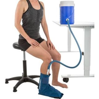
The initial phase of treatment often involves the RICE protocol: Rest, Ice, Compression, and Elevation.
Rest involves avoiding weight-bearing on the injured ankle. Ice packs can be applied for 15-20 minutes several times a day to help reduce swelling and pain.
Compression can be achieved using an elastic bandage to provide support and minimize swelling. Elevating the ankle above the level of the heart also helps to reduce swelling.
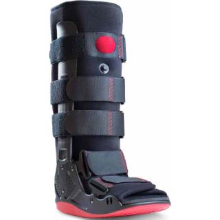
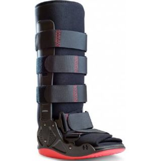
Immobilization is crucial in the early stages to allow the ligaments to heal. This may involve the use of a walking boot or a cast, depending on the severity of the sprain. The duration of immobilization can vary but is typically longer than that for a lateral ankle sprain, often ranging from several weeks to a few months.
Weight-bearing is usually restricted initially, with a gradual progression to bear weight as pain and swelling subside.
Physical therapy plays a vital role in the rehabilitation process. It focuses on restoring range of motion, improving strength in the surrounding muscles, enhancing proprioception (the body's awareness of its position in space), and gradually progressing back to activity.
Operative management is generally indicated for Grade III and some unstable Grade II high ankle sprains, as well as those associated with fractures. The goal of surgery is to anatomically reduce and stabilize the syndesmosis, allowing the ligaments to heal properly.
Several surgical techniques can be used:
Screw fixation involves placing one or more screws across the tibia and fibula above the level of the ankle joint to compress the bones together and stabilize the syndesmosis. These screws may be removed at a later stage once the ligaments have healed.
Another common surgical technique is suture button fixation. This involves placing strong sutures through small drill holes in the tibia and fibula and securing them with buttons on the medial and lateral sides of the lower leg. This allows for some physiological movement of the syndesmosis while still providing stability. Suture button fixation often allows for earlier weight-bearing compared to screw fixation and typically does not require a second surgery for removal.
The choice of surgical technique depends on the specific injury pattern and the surgeon's preference. Following surgery, a period of immobilization in a cast or boot is usually required, followed by a comprehensive rehabilitation program.
High Ankle Sprain Rehabilitation
Rehabilitation after a high ankle sprain is a crucial and often lengthy process. It is typically divided into several phases, each with specific goals:
The initial phase focuses on pain and swelling management. This involves continuing with the RICE protocol and gradually introducing gentle range of motion exercises, such as ankle pumps and alphabet tracing.
The second phase focuses on restoring range of motion. This includes more active range of motion exercises in all planes of movement, as well as stretching exercises to improve flexibility.
The third phase focuses on strengthening. This involves progressively loading the muscles around the ankle and lower leg, including the calf muscles, peroneal muscles, and tibialis anterior. Exercises may include calf raises, heel walks, toe walks, and resistance band exercises. The fourth phase focuses on proprioception and balance. This involves exercises that challenge the body's ability to maintain balance and control movement, such as single-leg stands, wobble board exercises, and agility drills.
The final phase involves a gradual return to sport or activity. This phase focuses on progressively increasing the intensity and duration of activities, ensuring that the ankle can tolerate the demands of the desired activity without pain or swelling.
The rehabilitation timeline for a high ankle sprain is typically longer than that for a lateral ankle sprain, often taking several months to a year for a full return to high-level athletic activity.
Despite appropriate treatment, some individuals may experience complications following a high ankle sprain. One potential complication is chronic syndesmotic instability, where the ligaments fail to heal properly, leading to persistent pain, weakness, and a feeling of giving way in the ankle. This may require further treatment, including possible revision surgery.
Another potential complication is persistent pain, even after the ligaments have healed. This may be due to nerve irritation, scar tissue formation, or other underlying issues. Post-traumatic arthritis can also develop in the ankle joint over time, particularly in cases of severe high ankle sprains or those associated with cartilage damage.
Preventing High Ankle Sprains
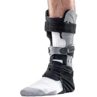
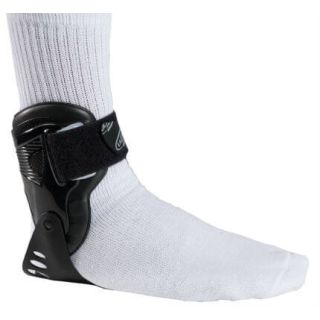
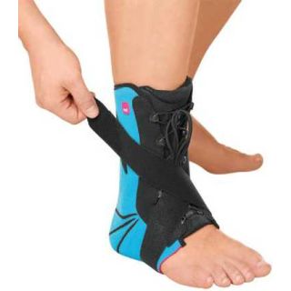
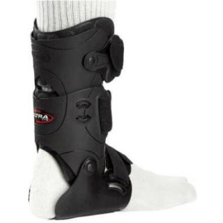
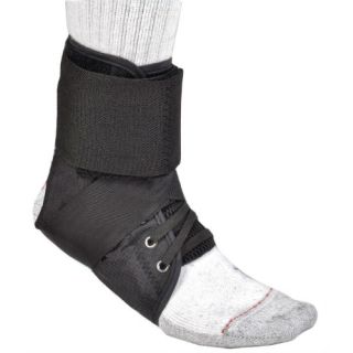
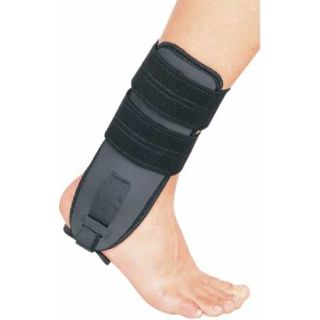
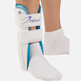
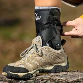
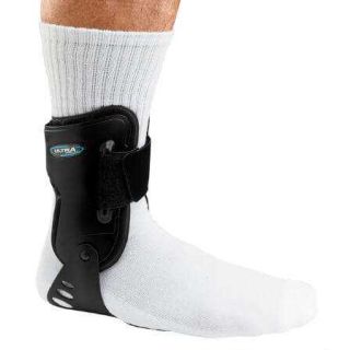
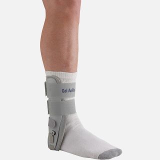
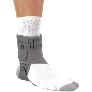
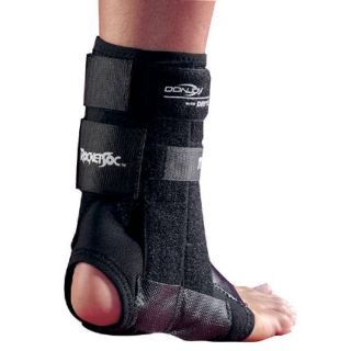
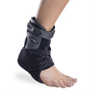
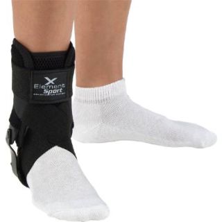
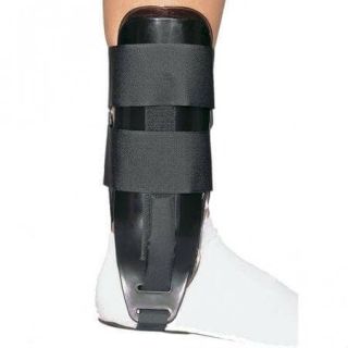
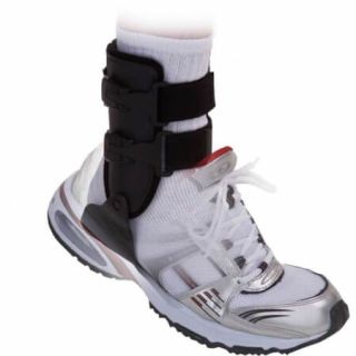
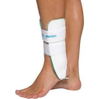
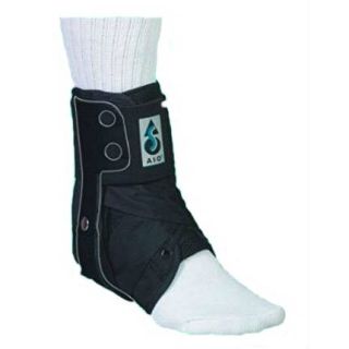
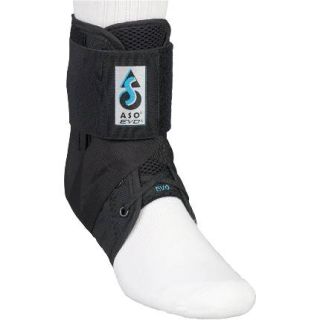
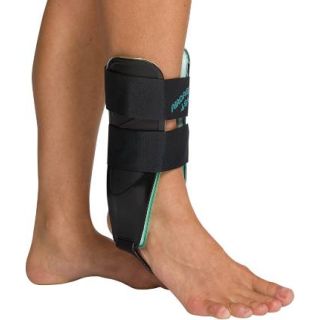
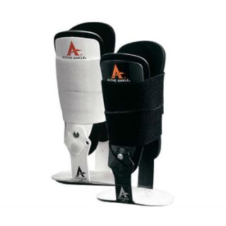
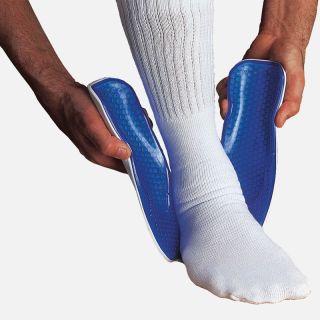
While it may not always be possible to prevent syndesmotic ankle sprains, there are some strategies that can help reduce the risk. These include ensuring proper warm-up before exercise, maintaining good strength and flexibility in the ankle and lower leg muscles, wearing appropriate footwear for the activity and/or a high ankle sprain brace/support, and avoiding sudden or excessive twisting movements of the ankle. Athletes participating in high-risk sports may benefit from taping or bracing of the ankle for added support.
Conclusion
In conclusion, the high ankle sprain, while less common than its lateral counterpart, represents a significant injury to the syndesmotic ligaments that connect the tibia and fibula.
Understanding the anatomy, mechanism of injury, diagnostic approaches, treatment options, and rehabilitation process is crucial for effective management and optimal recovery. Due to the complex nature of this injury and the potential for long-term complications, accurate diagnosis and appropriate treatment, often involving a prolonged rehabilitation period, are essential to ensure a successful return to activity and minimize the risk of future problems.
The insidious twist of a high ankle sprain demands a thorough understanding and a carefully tailored management plan to navigate the path back to full function.
Additional Resources:
Ankle support braces for injuries